Beranda
/ Leg Anatomy Muscles Ligaments And Tendons : leg aziz at Howard University College of Medicine - StudyBlue - Each muscle is connected to the corresponding bones to be moved via tendons.
Leg Anatomy Muscles Ligaments And Tendons : leg aziz at Howard University College of Medicine - StudyBlue - Each muscle is connected to the corresponding bones to be moved via tendons.
Insurance Gas/Electricity Loans Mortgage Attorney Lawyer Donate Conference Call Degree Credit Treatment Software Classes Recovery Trading Rehab Hosting Transfer Cord Blood Claim compensation mesothelioma mesothelioma attorney Houston car accident lawyer moreno valley can you sue a doctor for wrong diagnosis doctorate in security top online doctoral programs in business educational leadership doctoral programs online car accident doctor atlanta car accident doctor atlanta accident attorney rancho Cucamonga truck accident attorney san Antonio ONLINE BUSINESS DEGREE PROGRAMS ACCREDITED online accredited psychology degree masters degree in human resources online public administration masters degree online bitcoin merchant account bitcoin merchant services compare car insurance auto insurance troy mi seo explanation digital marketing degree floridaseo company fitness showrooms stamfordct how to work more efficiently seowordpress tips meaning of seo what is an seo what does an seo do what seo stands for best seotips google seo advice seo steps, The secure cloud-based platform for smart service delivery. Safelink is used by legal, professional and financial services to protect sensitive information, accelerate business processes and increase productivity. Use Safelink to collaborate securely with clients, colleagues and external parties. Safelink has a menu of workspace types with advanced features for dispute resolution, running deals and customised client portal creation. All data is encrypted (at rest and in transit and you retain your own encryption keys. Our titan security framework ensures your data is secure and you even have the option to choose your own data location from Channel Islands, London (UK), Dublin (EU), Australia.
Leg Anatomy Muscles Ligaments And Tendons : leg aziz at Howard University College of Medicine - StudyBlue - Each muscle is connected to the corresponding bones to be moved via tendons.. The muscles of the leg may be divided into three groups: They connect muscles to bones. Each muscle has tendons attached at each end. Muscles, either individually or in groups, are supported by fascia. The leg muscles are organized in 3 groups:
Anterior, lateral and posterior compartment. Learn how they work together to avoid injury and stay active. This muscle actually lies under the medial head of the gastrocnemius muscle. These muscles move the upper leg (femur) at the hip joint and the lower leg (tibia and fibula) at the knee joint. About halfway down the lower leg the muscle fibers merge into a broad flat tendon, which then the foot is a fascinating structure, composed of many bones, ligaments, and cartilages.
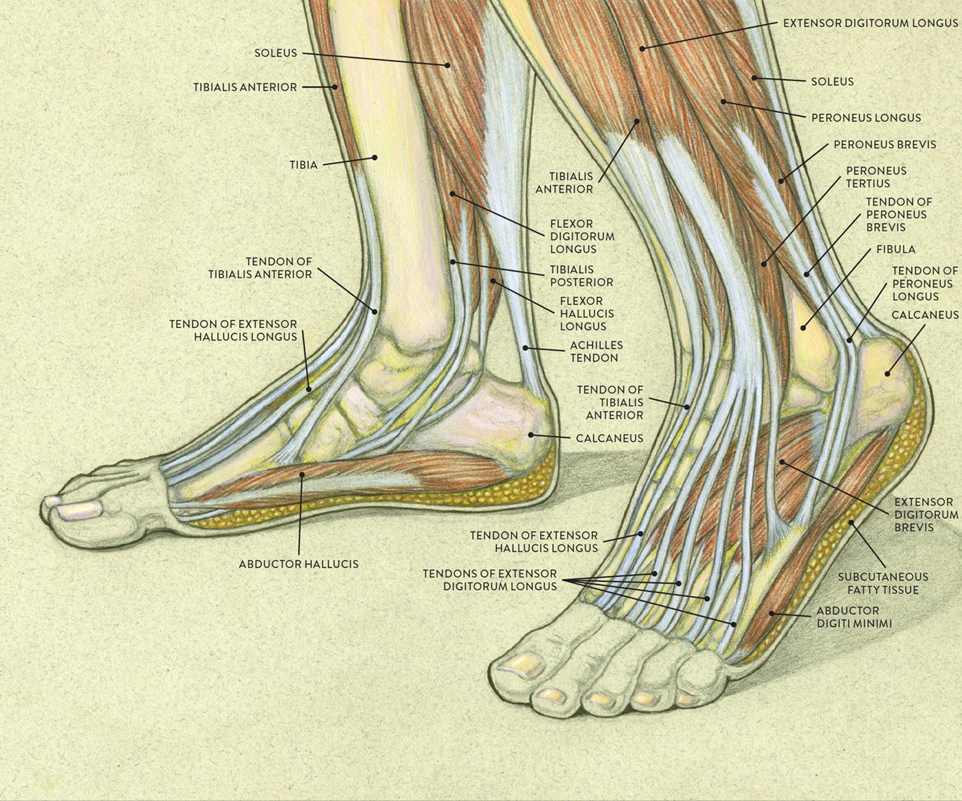
Foot Anatomy Tendons : Muscles Of The Foot Dorsal Plantar ... from doctorlib.info Each muscle is connected to the corresponding bones to be moved via tendons. The muscles of the leg may be divided into three groups: Tendons connect muscles to bones. Tendons consist of densely packed collagen fibers. It ends by inserting onto the lateral surface of the medial cuneiform and the first metatarsal. Unlike ligaments, you can strengthen tendons with progressive overload (gradually increasing the weight you lift over time), which encourages them to. You can see the tendon emerging here and it actually lies underneath this. There are four muscles in the anterior compartment of the leg.
, gustav andreisek2 and erika j.
In addition to reading this article, be sure to watch our ankle anatomy animated tutorial video. Tendons are designed to only stretch a small amount. Tendons and ligaments are bands of connective tissue that help stabilize the body and allow movement. Our bones are held together by ligaments and the bones are moved by muscles. There are four muscles in the anterior compartment of the leg. The leg muscles are organized in 3 groups: Tendons of the lower leg, muscles tendons and ligaments of the upper leg. 9.1 anatomy and normal mri appearance. Ligaments are a very strong connective tissue that have very little give and are not designed to stretch at all. Tendons consist of densely packed collagen fibers. The human leg, in the general word sense, is the entire lower limb of the human body, including the foot, thigh and even the hip or gluteal region. , gustav andreisek2 and erika j. One way our muscles work:
These all work together to bear weight. Anterior, lateral and posterior compartment. One way our muscles work: The muscle groups around the knee have an the muscles of the thigh and lower leg are comprised of compartments defined as distinct anatomical spaces bordered by fascia or bone. The leg muscles are organized in 3 groups:
IT Band Injury | Sports Podiatry from www.sportspodiatry.co.uk In other words, this page excludes information about the calf muscles… The individual bones are in turn connected by joints that are protected. Tendons are designed to only stretch a small amount. Tendons of the lower leg, muscles tendons and ligaments of the upper leg. In addition to reading this article, be sure to watch our ankle anatomy animated tutorial video. These all work together to bear weight. Originates from the lateral condyle of the tibia and the medial surface of the fibula. The cooperation of muscles, tendons and ligaments make our rigid skeleton a supporting and musculoskeletal system.
In other words, this page excludes information about the calf muscles…
Tendons and ligaments are bands of connective tissue that help stabilize the body and allow movement. 9.1 anatomy and normal mri appearance. The individual bones are in turn connected by joints that are protected. Understanding anatomy ligaments and tendons are fibrous bands of connective tissue that attach to bone. Collectively, they act to dorsiflex and invert the foot at the ankle joint. Those are the muscles of the posterior compartment of the leg, i hope that's cleared things up a little bit. The tendons of the edl can be palpated on the dorsal surface of the foot. Each muscle has tendons attached at each end. Tendons connect muscles to bones. The muscles, tendons, and ligaments that support the ankle joint work together to propel the body. Ligaments also support the lower end of the leg where it forms a hinge for the ankle. Other smaller muscles and tendons surround the knee joint as well. Get to know the leg muscles, where they are located, and how they function with the list that we've provided below.
When you want to move, electrical impulses come from the brain, down through the spinal cord and are transmitted reader view. The cooperation of muscles, tendons and ligaments make our rigid skeleton a supporting and musculoskeletal system. In addition, there are some other minor anatomical differences. The muscles, tendons, and ligaments that support the ankle joint work together to propel the body. They connect muscles to bones.
Conditions and Treatments from www.eorthopod.com About halfway down the lower leg the muscle fibers merge into a broad flat tendon, which then the foot is a fascinating structure, composed of many bones, ligaments, and cartilages. Ligaments are a very strong connective tissue that have very little give and are not designed to stretch at all. 12 photos of the muscles and tendons of the leg. Learn how they work together to avoid injury and stay active. Tendons connect muscles to bones. The tibialis anterior (tibialis anticus) is situated on the lateral side of the tibia; Learn about their differences and tendons connect muscles to bones, while ligaments connect bones to other bones. Tendons are tough bands of connective tissue found in the joints.
Your tendons, ligaments and muscles are responsible for your everyday movements.
In other words, this page excludes information about the calf muscles… There are four muscles in the anterior compartment of the leg. It is thick and fleshy above, tendinous below. Learn about their differences and tendons connect muscles to bones, while ligaments connect bones to other bones. Ligaments also support the lower end of the leg where it forms a hinge for the ankle. Learn about the muscles, tendons, bones, and ligaments that comprise the knee joint anatomy. The muscles of the leg may be divided into three groups: When the quadriceps muscles contract the patellar tendon is pulled and the leg straightens. Patellar tendon problems can arise from knee. Ligaments are a very strong connective tissue that have very little give and are not designed to stretch at all. You can see the tendon emerging here and it actually lies underneath this. The tibialis anterior (tibialis anticus) is situated on the lateral side of the tibia; Tendons are tough bands of connective tissue found in the joints.